Background: White matter hyperintensities (WMH) on T2-weighted MRI are common findings even in healthy elderly individuals. It is not known whether certain brain regions and arterial territories are particularly vulnerable to their development, and what the risk factors for WMHs are in otherwise healthy individuals.
Study Design: Cross-sectional MRI evaluation of a large random community sample as a sub-study of a study of ageing.
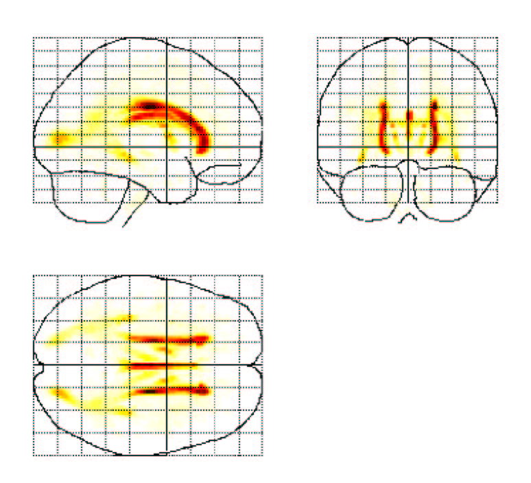
Methods: The study cohort comprised 2551 individuals, aged 60-64 years, recruited randomly from the electoral roll in Canberra. About one subject in five (N=478) was selected at random for participation in the MRI brain scan on a 1.5 T Philips scanner to obtain 1.5 mm thick T1-weighted (FSPGR) contiguous coronal sections and 4 mm thick T2-weighted FLAIR coronal slices through whole brain. Clinical (medical-psychiatric) and nutritional information was obtained, a brief cognitive assessment performed, and blood obtained for genetic and biochemical analyses. Whole brain WMH was extracted using a specially developed computer algorithm, and volumes of periventricular and deep brain WMHs obtained. WMHs were also partitioned into lobar regions and arterial territories after normalising each brain into Talairach space and developing a probability map using Statistical Probability Mapping (SPM99).
Results: WMHs are present in almost all brains, but their extent in this age group is small (median 0.5% of the white matter; range 0-8.5%). The periventricular regions are the most affected. Contrary to expectation, the frontal WM is not the most severely affected, and of the deep brain regions, the parieto-cccipital white matter bears the greatest load. There is significant hemispheric asymmetry of WMHs, which varies with gender. Female gender, hypertension and diabetes increase the propensity for WMH. Arterial territories were differentially affected.
Conclusions: This is the first detailed map of the topography of WMHs in brain of a representative sample and shows its wide spread and important determinants.
Back to S093 New Data on the Application of Translational Research to Patient Care
Back to The Eleventh International Congress